


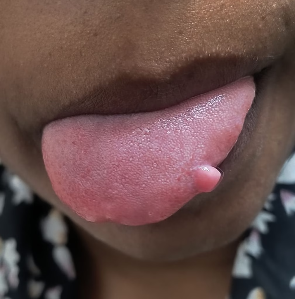
Quick answer: An irritation fibroma is a common, non-cancerous, scar-like lump that can form when the lining of the mouth is repeatedly rubbed or injured. It is often firm, smooth, painless, and similar in colour to the surrounding tissue. Because different oral conditions can look alike, a persistent mouth lump should be examined by a dentist or doctor rather than diagnosed at home.
What is an irritation fibroma?
An irritation fibroma—also called a traumatic fibroma or focal fibrous hyperplasia—is a reactive overgrowth of fibrous connective tissue. Despite the word “fibroma,” it is generally considered a reaction to long-standing irritation rather than a true tumour.
Repeated minor trauma can stimulate the tissue to produce dense collagen, creating a localised bump. The lesion itself is benign and does not turn into oral cancer. However, appearance alone cannot reliably distinguish every benign lump from other oral lesions, so professional assessment remains important.

What does an irritation fibroma look and feel like?
- Shape: usually a smooth, rounded or dome-shaped bump; it may have a broad base or sit on a short stalk.
- Texture: commonly firm or rubbery rather than fluid-filled.
- Colour: often matches the surrounding pink mucosa, but it can look paler, darker after bleeding, or ulcerated when repeatedly traumatised.
- Size: frequently around 1 cm or smaller, although larger lesions occur.
- Symptoms: usually painless; soreness can develop if it is bitten or rubbed.
- Growth pattern: commonly develops gradually over weeks or months and then remains stable.
The inner cheek along the biting line is the most common location. Irritation fibromas may also occur on the side of the tongue, inside the lower lip, gums, palate, or beneath a denture.


Common causes and sources of irritation
A source of repeated irritation is often present, although a person may not remember a specific injury. Common examples include:
- habitual cheek or lip biting;
- a rough, broken, or sharply positioned tooth;
- a rough filling, crown edge, or other restoration;
- rubbing from an ill-fitting denture or another dental appliance;
- repeated contact from orthodontic brackets or wires; or
- another persistent local mechanical irritant.
Plaque and calculus can contribute to some reactive gingival lesions, but poor oral hygiene alone should not be assumed to cause a fibroma. A clinician needs to examine the location and look for the most plausible irritant.
Is it dangerous or cancerous?
A confirmed irritation fibroma is benign and does not become cancerous. The safety concern is not transformation of the fibroma; it is the possibility of mistaking another condition for one. Mucoceles, papillomas, lipomas, giant cell fibromas, salivary-gland growths, vascular lesions, and some cancers can present as oral lumps.
A new mouth lump deserves prompt attention if it persists for about two weeks, enlarges, repeatedly bleeds, ulcerates, becomes hard or fixed, causes numbness or difficulty swallowing, or is accompanied by a neck lump or unexplained weight loss. These features do not automatically mean cancer, but they warrant timely examination.
How is irritation fibroma diagnosed?
A dentist, oral medicine specialist, oral surgeon, or other qualified clinician begins by asking when the lump appeared, whether it changes, and whether the area is repeatedly bitten or rubbed. The examination considers its size, colour, consistency, mobility, surface, exact location, and nearby teeth or appliances.
Why a biopsy may be recommended
Clinical findings may strongly suggest an irritation fibroma, but microscopic examination provides a definitive tissue diagnosis. A small lesion can often be completely removed as an excisional biopsy. A larger lesion, one in a sensitive location, or one with unusual features may instead need a smaller sample first.
The specimen is sent to a pathology laboratory to confirm the diagnosis and exclude look-alike conditions. Turnaround time varies between laboratories and may be longer if special testing is required, so the treating clinic should explain when and how results will be provided.
Treatment
An established fibroma is made of dense fibrous tissue and usually does not disappear simply with antibiotics, mouthwash, or topical gel. Treatment depends on diagnostic certainty, symptoms, size, and whether it interferes with chewing, speech, hygiene, or a dental appliance.
Remove or correct the irritant
The dentist may smooth a rough tooth or restoration, adjust a denture or orthodontic component, or help address a cheek-biting habit. Removing the source matters because continued trauma can cause the lesion to persist or recur after treatment.
Professional removal
When removal is appropriate, the area is usually numbed with local anaesthetic and the lesion is excised with a small margin. A scalpel, laser, or another surgical instrument may be used depending on the lesion, clinician, equipment, and need to preserve a suitable pathology specimen. No technique is best for every case.
Aftercare may include keeping the area clean, avoiding foods that irritate the wound, using pain relief recommended for the individual, and following instructions about stitches. Healing time and discomfort vary with the lesion’s size and location. Contact the treating clinic for bleeding that does not stop, increasing swelling, fever, pus, severe pain, or any other concern.
Can an irritation fibroma come back?
Recurrence is uncommon when the lesion is completely removed and the ongoing irritation is corrected. It can return if the source continues or if tissue remains. A recurrent or changing lump should be reassessed rather than repeatedly treated without confirming the diagnosis.
What not to do at home
- Do not cut, tie off, burn, or puncture the lump.
- Do not apply aspirin, corrosive chemicals, or unverified “removal” products.
- Do not take leftover antibiotics; a fibroma is not a bacterial infection.
- Do not postpone assessment because the lesion is painless.
While waiting for an appointment, avoid biting the area, choose softer foods if it is sore, and maintain gentle oral hygiene. Dental wax may temporarily reduce rubbing from an orthodontic wire, but the underlying cause still needs professional correction.
Frequently asked questions
Does an irritation fibroma go away by itself?
Established oral fibromas generally persist without treatment. Removing an irritant can prevent further trauma, but a remaining lump should be reviewed so the diagnosis and need for removal can be decided safely.
Is an irritation fibroma contagious?
No. It is a reactive tissue growth, not an infection, and it cannot spread from one person to another.
Can a fibroma be diagnosed from a photograph?
A photograph may help with triage, but it cannot confirm the diagnosis. Palpation, the clinical history, and sometimes biopsy are needed because multiple conditions can look similar.
Who should treat an oral fibroma?
A general dentist can assess the lesion and may treat a straightforward case. Referral to an oral medicine specialist, oral surgeon, periodontist, or another appropriate clinician may be advised when the diagnosis, location, size, or medical history makes the case more complex.
Key takeaway
An irritation fibroma is usually a harmless response to repeated local trauma, most often appearing as a firm, smooth, painless mouth bump. It does not turn into cancer, but self-diagnosis is unsafe because other lesions can resemble it. Arrange a dental or medical assessment for a persistent or changing oral lump, and never try to remove one yourself.
You may also find our guides to pyogenic granuloma and common dental problems helpful.
Reviewed by Dr. Manish Pandey, BDS. This article provides general dental education and does not replace diagnosis or personalised treatment. Any oral lump that persists, enlarges, ulcerates, bleeds, becomes painful or feels fixed should be examined promptly; biopsy may be needed to establish the diagnosis.