

Overview
Mastication (Chewing) is the first stage of digestion of food. Muscles of Mastication helps in grinding the food to make them small in size in the oral cavity.
Grinding the food is very important, it makes the food smaller in size. Smaller the size of the particle will increase the surface area. This food can then be swallowed with ease and further chymified with the help of enzymes as it moves along the alimentary canal.
Many different structures like Teeth, Bone (Mandible and maxilla) and muscles like tongue and masticatory muscles work together to enable a person to chew the food.
Embryologically, the muscles of mastication are developed from the first pharyngeal arch.

Primary Muscles Of Mastication
There are many muscles which help in mastication. First, we will discuss primary muscles which are as follow:
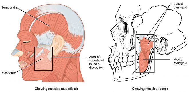
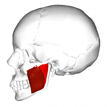
- Masseter
- Temporalis
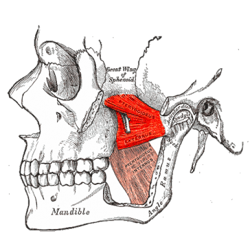
- Medial pterygoid
- Lateral pterygoid
Accessory Muscles Of Mastication
There are some other muscles that help in mastication. Their names are as follows.
Suprahyoid Muscles
- Mylohyoid
- Geniohyoid
- Stylohyoid
- Digastric
Infrahyoid muscles
- Thyrohyoid
- Omohyoid
Now, in this article, we will know about the Anatomy, attachment innervation and action of these muscles individually.
Masseter Muscle
It is the strongest in all muscles. Masseter muscle is quadrangular in shape, and it can be divided into two parts: Deep and Superficial.

Origin
- Superficial layer: Tendinous aponeurosis from the zygomatic process of the maxilla and anterior 2/3 of the inferior border of the zygomatic arch.
- Deep layer: Medial aspect and inferior border of posterior one-third of the zygomatic arch
Insertion
- Superficial Part: Lower border of the lateral surface of the mandible.
- Deep Layer: Upper part of the ramus and coronoid process.
Functions of Masseter Muscle
- The masseter muscle functions as a powerful elevator of the jaw.
- Lateral movement of the mandible
- Retraction of mandible
Nerve Supply
The masseter muscle is innervated by Messetric Nerve derived from the mandibular division of the Trigeminal nerve.
Blood supply
Vascular supply from the masseteric branch of the maxillary artery.
Temporalis Muscle
Temporalis Muscle is the fan-shaped muscles originated from temporal fossa of the skull. It is a large muscle which covers the temporal bone of the skull. The primary function of this muscle is to elevate the mandible.

Origin of Temporalis Muscle
The inferior temporal line across the entire temporal fossa, including parts of the parietal and most of the squama of the temporal bones.
Insertion
As a tendon on Coronoid Process of the mandible.
Blood supply of Temporalis Muscle
Vascular supply of the Temporalis Muscle is via branches of the superficial temporal and maxillary arteries.
Nerve Supply
Anterior and posterior deep Temporal nerve from mandibular division of trigeminal nerve.
Function
- Elevation of mandible
- Retraction of the mandible: by Posterior and middle portion of the muscle.
Lateral Pterygoid Muscle
The Lateral Pterygoid Muscle has two heads of origin, smaller Superior head, and larger inferior head.

Origin
- Superior Head: Infratemporal region of the greater wing of the sphenoid bone.
- Inferior Head surface of the lateral pterygoid plate.
Insertion
At the neck of the mandible and various structures of the TMJ.
Nerve Supply
Branch of the buccal nerve from the mandibular division of the trigeminal
nerve.
Blood Supply
Pterygoid branch of the maxillary artery.
Function
- Depresses the mandible so, It is also called a jaw opener muscle.
- Protrudes forward for opening the jaw.
- It also helps to side movement of the mandible.
Medial Pterygoid Muscle
The Medial Pterygoid Muscle is the deepest situated muscle of mastication.

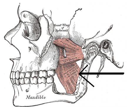
Origin
Deep head of lateral pterygoid plate and maxillary tuberosity
Insertion
In the medial angle of the mandible
Nerve Supply
Mandibular division of the trigeminal nerve
Blood Supply
Pterygoid branch of the 2nd part of maxillary artery
Function
- Elevates the mandible
- Closes the jaw
- Side to side movement
Accessory Masticatory Muscles
Digastric
- Two bellies unite by tendon
- Act as a depressor muscle adding to the action of lateral pterygoid muscle
Mylohyoid
- It is a flat triangular muscle.
- A secondary role in mastication as a depressor of the mandible.
- Mylohyoid muscle elevates the floor of the mouth to help in deglutition.
Geniohyoid
- Short and narrow muscle which lies over mylohyoid muscle.
- It depresses the mandible.