Overview
Ameloblastoma is a rare, noncancerous tumor of the jaw. Adamantinoma and Adamantoblastoma are the synonyms of this tumor. The name is derived from Amel meaning teeth and Blastos meaning germ. This tumor is frequently seen in the lower jaw, close to wisdom tooth or molar.
What causes Ameloblastoma?
The causes of this tumor are not clear. The causes may include injury to the teeth or jaw, infection of gum and teeth. Infection of the virus and low protein in the person’s diet may play a major role in the development of this tumor((https://rarediseases.org/rare-diseases/ameloblastoma/)).
Symptoms
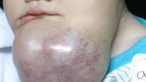
This is a slow-growing tumor of the jaw. It is found in both Maxilla and Mandible. However, up to 80% it has been seen in Mandible only. It is slow growing painless swelling which leads to facial deformity.
Due to the increase in the size of the tumor, the teeth become loose. There will also be malocclusion because of the movement of the tooth.

The lesion causes expansion of bony carticle plates. Thin shells of bone will be formed ahead of the lesion. This shell of bone will crack on palpation. This phenomenon is called as “Egg Shell Cracking” or Crepitus.
In some cases, there will not be any presentable symptom. They diagnosed accidentally when they come for another treatment.
Sometimes, it grows very quickly with pain. It can spread to the eyes, nasal sinus and skull.
Very rarely, ameloblastoma cells can reach other areas of the body such as lymph nodes in the neck and lungs.
How common is Ameloblastoma?
Ameloblastoma is a rare slow growing but a locally invasive tumor. In the United States, between 300 and 600 cases are diagnosed each year. It accounts for 1% of all oral tumors and almost 9-11% of all odontogenic tumors((https://www.ncbi.nlm.nih.gov/pmc/articles/PMC4439660/)). It is equally common in men and women at any age, but usually, it is seen between 30 to 40 years of age.((https://www.ncbi.nlm.nih.gov/pmc/articles/PMC4439660/))
There is conflicting proof of the incidence rates in different races. Although some reports claim an increased incidence of ameloblastoma in black people ((Shafer’s Textbook of Oral Pathology – Rajendran, R, Sivapathasundaram, B))
Types
If we discuss the types of tumors, they are classified based on their histopathology and clinical characteristics.
Histopathology
- Follicular
- Acanthomatous
- Granular cell
- Basal cell
- Desmoplastic
- Plexiform
Clinical Features
- Central/Intraosseous
- Conventional/Multicystic/solid (Most common)
- Unicystic
- Peripheral (Extraosseous)
- Pituitary Ameloblastoma
- Malignant Ameloblastoma (Rare)
Ameloblastoma Radiographic features
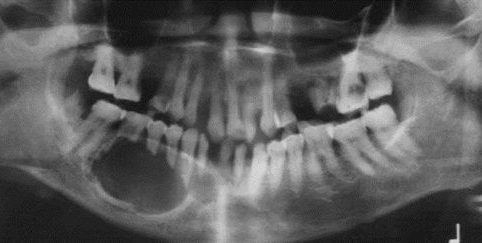
In the radiograph, tumor area appears as a rounded well-defined margin in the bone with varying size and features. Multiple round radiolucencies can be seen in the larger (multilocular) tumor. It gives a characteristic “soap bubble” appearance. A single round radiolucency can be seen on the unicystic tumor radiograph.
Treatment
Surgery to get rid of the tumor
Ameloblastoma treatment usually includes surgery to remove the neoplasm. Ameloblastoma typically grows into the nearby jawbone, therefore surgeons may need to remove the affected part of the jawbone. an aggressive approach to surgery reduces the chance that ameloblastoma will return.
Surgery to repair the jaw
If surgery involves removing a part of your jawbone, surgeons will repair and reconstruct the jaw. this will facilitate improve how your jaw looks and works subsequently. The surgery can also assist you to be able to eat and speak.
Radiation therapy
Radiation therapy using high-powered energy beams may well be required when surgery or if surgery is not a choice.
Supportive care
A variety of specialists will assist you to work through speaking, swallowing and eating problems throughout and after treatment.
[…] Causes Types Radiographic Features and Treatment Ameloblastoma: Symptoms Causes and Radiographic feature of Jaw tumor 8 Most Common Dental Problems Dental Veneers: Everything you need to know Your kids needs a 6 […]