Overview
In true sense, The term Odontoma refers to any tumor of odontogenic origin. Odontoma is considered more as a hamartomatous malformation,
also as a composite lesion, as it contains more than one tissue.
Causes of Odontoma
It is one of the most common non-cancerous tumors of Odontogenic origin. Formation of Odontoma is due to the irregular growth of epithelial and ectomesenchymal cells during tooth formation.
This is considered more like a composite lesion or hamartomatous formation, as it contains more than one type of the tissue.
The etiology of this tumor is still unknown. It may be related to various pathological conditions like local trauma, inflammatory and/or infectious processes or maybe because of hereditary conditions. ((https://www.ncbi.nlm.nih.gov/pmc/articles/PMC5034075/))
Types of Odontoma
Histopathologically and Radio-graphically it is recognizable in two forms:
- Compound Odontome – Toothlike structure may resemble teeth. More common in Maxilla
- Complex Odontome – Consists of all tooth forming structure but in a disorderly pattern. Calcified tissues found as irregular masses without any similarity to morphology or teeth. More common in Mandible (67 percent). ((Textbook of Oral and Maxillofacial Surgery 3rd edition by Prof. Dr. Neelima Anil Malik))
According to WHO Odontome can be classified as follows
- Ameloblastic fibro-odontome – Consists of varying amounts of calcified dental tissue
- Odonto-ameloblastoma – It is a very rare tumor and represents as ameloblastoma in clinical and structural form but it contains Enamel and dentin.
- Complex odontome –Here the calcified dental tissues arranged in irregular form without any morphological similarity to teeth.
- Compound odontome-It consists of formed calcified toothlike structures or miniature dwarfed teeth.
Radiographic Features Of Odontoma
Complex and Compound odontoma presents different radiographic features:
Complex
It may appear small large or occasionally huge irregular radio-opaque mass. Complex odontome often surrounded by a thin radiolucent zone. This is frequently associated with a displaced unerupted tooth.
Initially, it presents complete radiolucency, as it calcifies it also presents with radiopacity. In some cases, it can present mixed radiolucency and radiopacity.
Compound
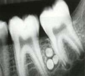
Appears as a radiopaque mass of calcified structure which resembles with the tooth. This tooth like radioopaque images may be smaller than normal tooth. This radiopacity may be surrounded by a radiolucent zone. Sometimes it prevents the eruption of permanent teeth.
Treatment
Completely calcified complex or compound odontoma can be left inside as it is bio-inert. Surgical removal may be necessary if it blocks the tooth eruption. Recurrence of odontoma is not seen. Surgical excision may also be needed for definite diagnosis.